Research Focus
We develop next-generation MRI methods to uncover how cells are built, how they function, and how they change in health and disease. Our work goes beyond static images—toward dynamic, quantitative descriptions of cellular architecture, composition, and physiology.
At the core is the idea that MRI can probe the microscopic world of cells through motion. By combining advanced diffusion and flow-sensitive measurements, we capture how water and metabolites move within complex cellular environments. These motions are not random—they are shaped by membranes, organelles, macromolecules, and barriers—turning MRI into a powerful, non-invasive window on cellular structure and function.
Leveraging the enhanced spectral and spatial resolution of ultra-high field MRI, we push sensitivity to new limits, enabling us to disentangle subtle contributions from tissue chemistry, compartmental exchange, and microvascular flow. While diffusion MRI is traditionally framed as a tool for mapping geometry—cell size, shape, and orientation—we treat this as only part of the story.
Cellular systems are governed not just by structure, but by interactions: chemical composition, permeability, fluid dynamics, and the integrity of biological barriers. These factors leave measurable fingerprints in MR signals. By rethinking experimental design and modeling, we aim to decode these signals to reveal deeper physiological insights—linking microstructure to metabolism, transport, and mechanical properties.
Our vision is to transform MRI from a descriptive imaging modality into a quantitative tool for cellular biophysics in vivo, bridging scales from molecules to tissue and enabling new biomarkers of disease and treatment response.
Research projects
C-MORPH
C-MORPH develops advanced MRI methods to map cell-type specific properties in the living brain and spinal cord. By combining diffusion encoding, spectroscopy, and novel imaging, we disentangle microstructural and pathological processes that are otherwise entangled in conventional scans. The goal is to deliver sensitive, clinically applicable markers of disease progression, inflammation, and neurodegeneration for personalized medicine.
Software
Code for experimental design of diffusion MRI experiments with general gradient waveforms https://github.com/samo-lasic/Lasic_SPAS_ImagingNeuroscience2025
Selected Group Publications
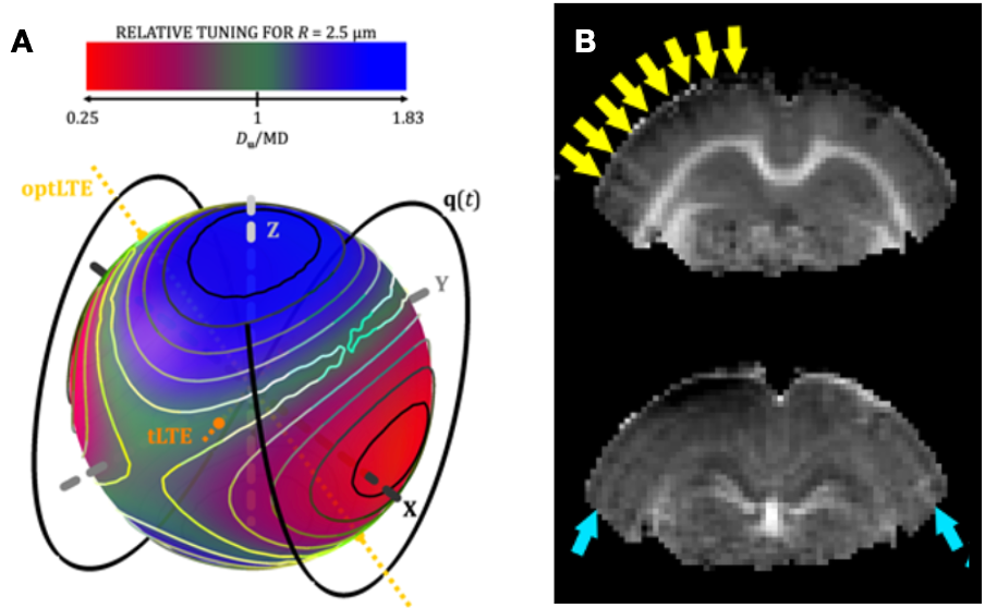
Samo Lasič, Nathalie Just, Markus Nilsson, Filip Szczepankiewicz, Matthew Budde, Henrik Lundell (2025) Spectral Principal Axis System (SPAS) and tuning of tensor-valued encoding for microscopic anisotropy and time-dependent diffusion in the rat brain, Imaging Neuroscience 3: IMAG.a.35
M Jokivuolle, F Mahmood, KH Madsen, FSG Harbo, L Johnsen, H Lundell (2025) Assessing tumor microstructure with time-dependent diffusion imaging: Considerations and feasibility on clinical MRI and MRI-Linac, Medical Physics 52(1):346-361
S Lasič, A Chakwizira, H Lundell, CF Westin, M Nilsson (2024) Tuned exchange imaging: Can the filter exchange imaging pulse sequence be adapted for applications with thin slices and restricted diffusion? NMR in Biomedicine 37 (11), e5208
Lasič S, Yuldasheva N, Szczepankiewicz F, Nilsson M, Budde M, Dall’Armellina E, Schneider JE, Teh I, Lundell H (2022) Stay on the beat with tensor-valued encoding: time-dependent diffusion and cell size estimation in ex vivo heart, Frontiers in Physics 2022:812115
Lundell H, Najac C, Bulk M, Kan HE, Webb A G, Ronen I (2021) Compartmental diffusion and microstructural properties of human brain gray and white matter studied with double diffusion encoding magnetic resonance spectroscopy of metabolites and wate, Neuroimage, 234:117981
Henriques RN, Palombo M, Jespersen SN, Shemesh N, Lundell H, Ianuş A (2020) Double diffusion encoding and applications for biomedical imaging, Journal of Neuroscience Methods, 108989
Andersen KW, Lasič S, Lundell H, Nilsson M, Topgaard D, Szczepankiewicz F, Siebner HR, Blinkenberg M, Dyrby TB (2020) Disentangling white-matter damage from physiological fiber orientation dispersion in multiple sclerosis, Brain Communications, 2:fcaa077
Lundell H, Ingo C, Dyrby TB, Ronen I (2020) Cytosolic diffusivity and microscopic anisotropy of N‐acetyl aspartate in human white matter with diffusion‐weighted MRS at 7 T, NMR in Biomedicine, e4304
Lundell H, Nilsson M, Dyrby TB, Parker GJM, Hubbard Cristinacce PL, Zhou F-L, Topgaard D, Lasič S, (2019) Multidimensional diffusion MRI with spectrally modulated gradients reveals unprecedented microstructural detail, Scientific Reports 9:9026
Scharff Nielsen J, Dyrby TB, Lundell H. (2018) Magnetic resonance temporal diffusion tensor spectroscopy of disordered anisotropic tissue. Scientific Reports 8:2930
Collaborators: updated Itamar Ronen, Brighton, UK Faisal Mahmood, SDU, Odense, Denmark Lydianne Hirschler and Thijs van Osch, LUMC, Leiden, The Netherlands Markus Nilsson, LU, Lund, Sweden Christoffer Steele, Concordia University, Montreal, Canada Rodrigo Moreno, KTH, Stockholm, Sweden