Cortical lessions and grey matter dysfunction in Multiple Sclerosis
Imaging plays a pivotal role in multiple sclerosis (MS) diagnosis and monitoring. It can help us uncover and understand the yet unknown mechanisms that contribute to the large variety of symptoms in MS.
For example, cortical pathology is now known to significantly contribute to disability in multiple sclerosis (MS) and has therefore attracted considerable interest in the last decade. However, we are yet to understand the impact of cortical damage on both the connectivity and functional integrity of the affected area and other parts of the central nervous system.
In this project, we exploit the greater sensitivity of 7 Tesla magnetic resonance imaging (MRI) to cortical lesions, together with quantitative imaging sequences and transcranial magnetic stimulation (TMS) to comprehensively map the role of cortical damage in different subtypes of MS.
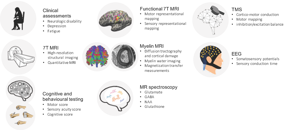
Overview of the experimental protocol and methodology used in the project.
Using both MRI and TMS, this project addresses two main research questions:
- What is the impact of individual cortical lesions, specifically in the primary sensorimotor hand area, on hand dexterity in different subtypes of MS
- What role do focal and diffuse cortical myelin changes play in relation to physical and cognitive function in primary progressive MS
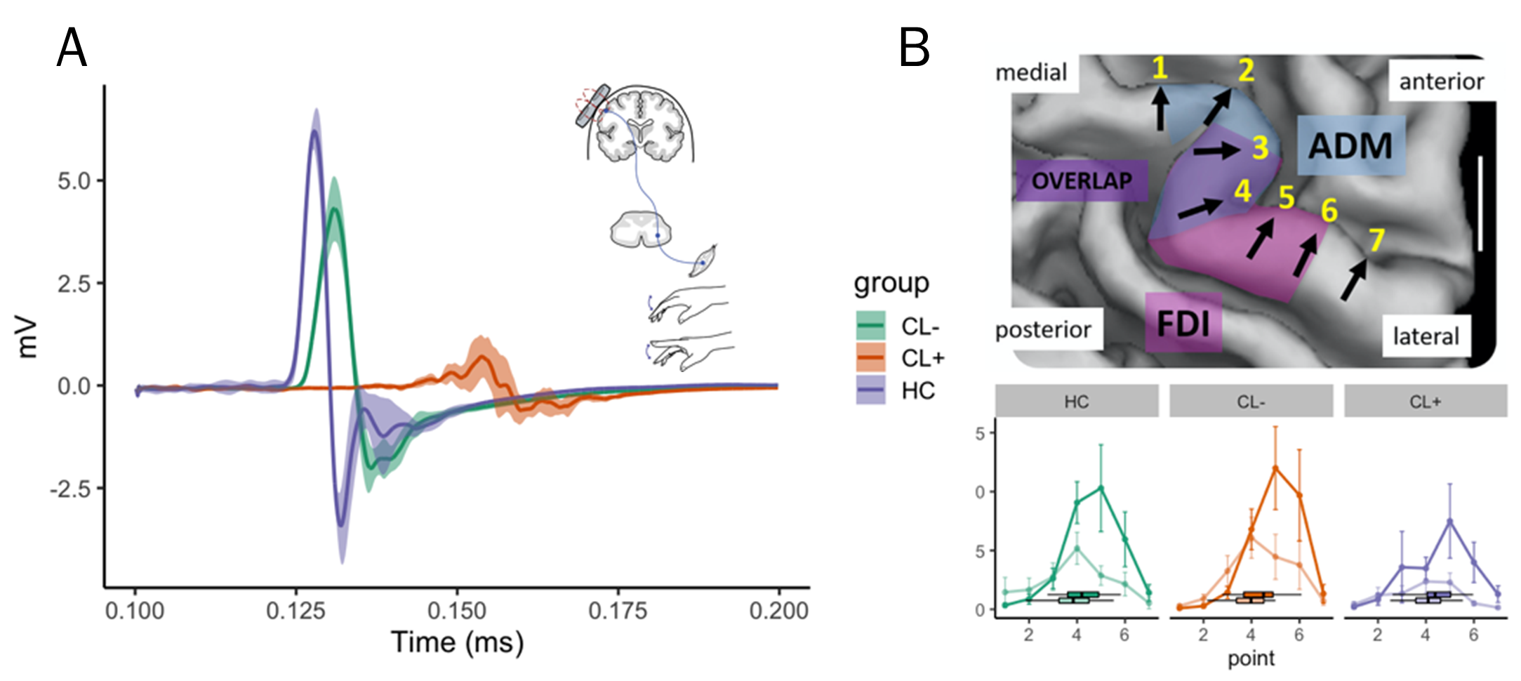
(A) The three motor evoked potentials (MEP) traces show that the onset latency was longer and lower in amplitude for both relapsing remitting MS patients compared to a healthy control (HC), but much more so in the patient with cortical damage (CL+). (B) TMS motor mapping successfully separates the representation of the FDI (bold) and ADM (faded color) muscle in all three groups.
Impact
This project will reveal key insights into how cortical demyelination and damage, both regionally and globally, contribute to cognition and motor impairment in MS, two major disabling problems for patients. We will advance the possibilities of MRI to capture cortical involvement in Danish MS patients with the goal to improve individual stratification, monitoring of disease progression and capturing of the individual response to therapy.
Selected Publications
[f31547f2-5d3f-415e-8e43-bb2962756b69 23674430-9575-4356-8cad-3909639392ce 81ee3951-9c26-43db-a9d4-7ba901789c32 5c799920-9730-4648-a81a-a5e495e6d7dc 4a146737-dd7b-43b2-b9e8-a3912c32e144 3e22a5a7-71ae-43cb-8621-cb6b10dc3253 7216e9d3-92e5-4342-a874-e408b042f132 986e451e-ee82-441b-a93a-3ae4259a024b 6c52d5a2-aee5-4da2-bac8-ec2395b27cdd 68cdf529-7839-4a8a-bc8a-37171142a0ca eb47f1d1-f66f-4e2e-95bb-fcd9bf6d9e32 e4cf28fd-6895-45e3-ba48-018381644ad4 620c0cf0-2e1c-4a0e-8a15-7ea0f13c0ed8 07dbffd2-79d9-43a4-ab98-be7a97d50d09 aca28f0a-ecf9-41d5-b801-90ee952ceb8b e8e401c0-a025-4875-83d5-89141dd82933 c71ae103-3f09-4ce5-a303-9bf100798831 d7014ac4-f9ce-407a-ba74-64be3c0675d1 92c86220-edd1-47c8-b887-6d3843bf85a8 31250f6c-30ea-4105-9d59-67b5f59fc8d9 c8bdc1b3-d5d7-4cae-a823-353efa20332e f1858b1b-99eb-4da9-b1ac-bfa1e6056d39 cc26fe3c-0f94-4066-a19c-4d91931953aa 7dd4359e-311a-4ae8-b0ea-8811bc051672 07733d3a-df55-4cbc-bbfa-d3dc04670347 e7e4cdd0-f078-4f29-beec-5eec92165706 2fb50b4f-0a2e-489a-bffa-3e2c829d7154 3c687e7f-5a39-4695-b82d-8ec29c45bd9a e4dd1532-1969-4477-af6d-c8746d2f4851 fe99a59d-3287-437d-895f-709750b7728b 63c80110-4e06-4031-a357-0097f3b30f75 63e63839-2779-495d-ac20-cd9c4dcf4d64 f05e524a-216e-474b-a44d-e720af9e97a1 ff8d1f54-f75e-44f3-9c1a-5bd2871e811b 3447021f-c53b-45e8-9179-de0819d6a0bc c28b70a6-2e8f-4499-ab68-1d0a4c463840 5ad33576-0e7f-4860-a215-516c0209f1e8 8a52b608-0dce-4536-ba75-1082b002d3ec 7f3f836f-96b9-4583-9e51-d5757dcf104c 3676fbde-ce2e-4828-9b0e-1708e46ad8f3 7176c03f-1fa1-46da-97a8-b2e3af7e330d d263ef6d-c108-4b8f-ba52-41660a5c8fd2 24f39aad-dc06-4c68-a20a-1deed570c1ac 4c294409-c0e1-44fd-889e-9055720e5483 f865d1b2-1137-4472-be9c-a6e89f4e7628 ee1559c8-81ed-4f75-98e8-26185560db73 89e0359b-1ea7-4708-8000-145922b4ca5a 25858831-1dd1-4cd5-ad29-ab86184fbf25 1ac3462e-2790-4046-ac36-5a54fd41eb7c c677afed-1530-4763-a18b-bd0deb7d44a2 0ed286cd-930f-4026-81ff-4ba9c3507457 ec2ccaa6-aefa-45b6-a330-77a469fdd20b 238a20d2-1344-413a-adee-e0d3d77ba727 d3330024-288c-4ff9-8608-06df2fab0ea3 91668e02-3932-4c91-975f-01f654728a0d dd747b0d-0d73-4a1e-981a-36fa97f68f03 2dacc43f-6928-403a-9db5-d67256fc3803 5d6f7443-3b76-4026-83b0-4da8a8984212 9d11c127-1fc5-4037-aae5-cd1cc460c0c3 5c314301-e9aa-4ea6-b293-5a5b423055d7 a50b88ea-9490-41a9-bb53-001eab673228]
Madsen MAJ,
Wiggermann V,
Marques MFM,
Lundell H,
Cerri S,
Puonti O,
Blinkenberg M,
Christensen JR,
Sellebjerg F &
Siebner HR, Linking lesions in sensorimotor cortex to contralateral hand function in multiple sclerosis: a 7 T MRI study,
[map[#text:Brain -lang:en] map[#text:Brain -lang:en] map[#text:Brain : a journal of neurology -lang:en]]
vol 145,
Issue 10,
p. 3522-3535,
(2022).
DOI:10.1093/brain/awac203
[f31547f2-5d3f-415e-8e43-bb2962756b69 23674430-9575-4356-8cad-3909639392ce 81ee3951-9c26-43db-a9d4-7ba901789c32 5c799920-9730-4648-a81a-a5e495e6d7dc 4a146737-dd7b-43b2-b9e8-a3912c32e144 3e22a5a7-71ae-43cb-8621-cb6b10dc3253 7216e9d3-92e5-4342-a874-e408b042f132 986e451e-ee82-441b-a93a-3ae4259a024b 6c52d5a2-aee5-4da2-bac8-ec2395b27cdd 68cdf529-7839-4a8a-bc8a-37171142a0ca eb47f1d1-f66f-4e2e-95bb-fcd9bf6d9e32 e4cf28fd-6895-45e3-ba48-018381644ad4 620c0cf0-2e1c-4a0e-8a15-7ea0f13c0ed8 07dbffd2-79d9-43a4-ab98-be7a97d50d09 aca28f0a-ecf9-41d5-b801-90ee952ceb8b e8e401c0-a025-4875-83d5-89141dd82933 c71ae103-3f09-4ce5-a303-9bf100798831 d7014ac4-f9ce-407a-ba74-64be3c0675d1 92c86220-edd1-47c8-b887-6d3843bf85a8 31250f6c-30ea-4105-9d59-67b5f59fc8d9 c8bdc1b3-d5d7-4cae-a823-353efa20332e f1858b1b-99eb-4da9-b1ac-bfa1e6056d39 cc26fe3c-0f94-4066-a19c-4d91931953aa 7dd4359e-311a-4ae8-b0ea-8811bc051672 07733d3a-df55-4cbc-bbfa-d3dc04670347 e7e4cdd0-f078-4f29-beec-5eec92165706 2fb50b4f-0a2e-489a-bffa-3e2c829d7154 3c687e7f-5a39-4695-b82d-8ec29c45bd9a e4dd1532-1969-4477-af6d-c8746d2f4851 fe99a59d-3287-437d-895f-709750b7728b 63c80110-4e06-4031-a357-0097f3b30f75 63e63839-2779-495d-ac20-cd9c4dcf4d64 f05e524a-216e-474b-a44d-e720af9e97a1 ff8d1f54-f75e-44f3-9c1a-5bd2871e811b 3447021f-c53b-45e8-9179-de0819d6a0bc c28b70a6-2e8f-4499-ab68-1d0a4c463840 5ad33576-0e7f-4860-a215-516c0209f1e8 8a52b608-0dce-4536-ba75-1082b002d3ec 7f3f836f-96b9-4583-9e51-d5757dcf104c 3676fbde-ce2e-4828-9b0e-1708e46ad8f3 7176c03f-1fa1-46da-97a8-b2e3af7e330d d263ef6d-c108-4b8f-ba52-41660a5c8fd2 24f39aad-dc06-4c68-a20a-1deed570c1ac 4c294409-c0e1-44fd-889e-9055720e5483 f865d1b2-1137-4472-be9c-a6e89f4e7628 ee1559c8-81ed-4f75-98e8-26185560db73 89e0359b-1ea7-4708-8000-145922b4ca5a 25858831-1dd1-4cd5-ad29-ab86184fbf25 1ac3462e-2790-4046-ac36-5a54fd41eb7c c677afed-1530-4763-a18b-bd0deb7d44a2 0ed286cd-930f-4026-81ff-4ba9c3507457 ec2ccaa6-aefa-45b6-a330-77a469fdd20b 238a20d2-1344-413a-adee-e0d3d77ba727 d3330024-288c-4ff9-8608-06df2fab0ea3 91668e02-3932-4c91-975f-01f654728a0d dd747b0d-0d73-4a1e-981a-36fa97f68f03 2dacc43f-6928-403a-9db5-d67256fc3803 5d6f7443-3b76-4026-83b0-4da8a8984212 9d11c127-1fc5-4037-aae5-cd1cc460c0c3 5c314301-e9aa-4ea6-b293-5a5b423055d7 a50b88ea-9490-41a9-bb53-001eab673228]
Madsen MAJ,
Wiggermann V,
Bramow S,
Christensen JR,
Sellebjerg F &
Siebner HR, Imaging cortical multiple sclerosis lesions with ultra-high field MRI
[map[#text:NeuroImage: Clinical -lang:en] map[#text:NeuroImage. Clinical -lang:en]]
vol 32,
p. 1-14,
(2021).
DOI:10.1016/j.nicl.2021.102847
Madsen MAJ, Povazan M, Wiggermann V, Lundell H, Blinkenberg M, Romme Christensen J, Sellebjerg F, Siebner HR. 2024. Association of cortical lesions with regional glutamate, GABA, N-Acetylaspartate, and Myoinositol levels in patients with multiple sclerosis. Neurology. 103(1): e209543. https://doi.org/10.1212/WNL.0000000000209543
Madsen MAJ, Wiggermann V, Marques MFM, Lundell H, Cerri S, Puonti O, Blinkenberg M, Romme Christensen J, Sellebjerg F, Siebner HR. Linking lesions in sensorimotor cortex to contralateral hand function in multiple sclerosis: A 7 T MRI study. Brain, 145(10), 2022, DOI: 10.1093/awac203
Madsen MAJ, Wiggermann V, Bramow S, Romme Christensen J, Sellebjerg F, Siebner HR. Imaging cortical multiple sclerosis lesions with ultra-high field MRI. NeuroImage: Clinical, 32, 2021, DOI:10.1016/j.nicl.2021.102847