Research Focus
The Movement Disorders group bridges clinical, computational and cognitive neuroscience in combination with brain stimulation to advance the pathophysiological understanding of movement disorders.
The Movement Disorders group is headed by David Meder. Our research primarily focuses on Parkinson´s disease (PD), but also REM-sleep behavior disorder and progressive supranuclear palsy.
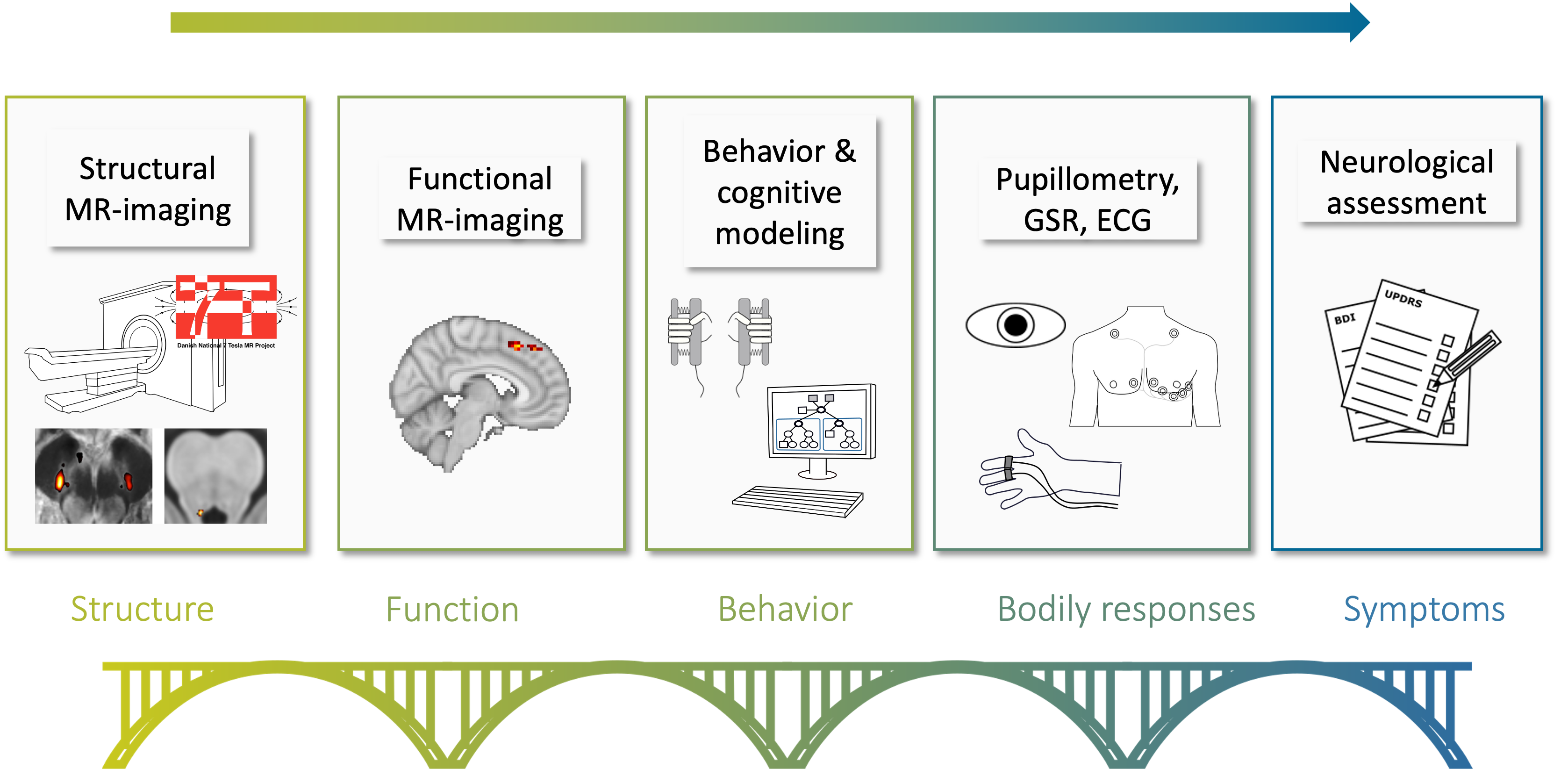
The mission of the Movement Disorders group is to use chemogenetic and optogenetic animal models together with advanced brain mapping techniques in humans, combining them with computational modeling to investigate how movement disorders alter brain function and structure in motor, cognitive and limbic systems. We use the ultra-high field (7 tesla) MR scanner in order to map the structural integrity of midbrain nuclei at high resolution. This allows us to investigate the relationship between the individual spatial pattern of neurodegeneration in Parkinson’s disease, the resulting changes in brain function and finally the patient’s clinical symptoms.
We apply computational models of learning and decision-making to probe disease-induced changes in brain and behavior. Traditionally, clinical neuroimaging has often taken a predominantly descriptive approach, describing that there is a change in brain or behavior observed in the disorder. Computational neurology goes one step further: We create computer algorithms that mimic how the function under investigation might be solved by the brain. If such a model then fits to the observed behavior and neural activity, we can infer that the brain (approximately) uses the kind of algorithm we defined in the model. Observing which parameters of the model are changed in the disease can then lead to a mechanistic understanding, explaining not only that there are disease-induced changes, but how the changes occur at the neural and symptomatic level.
Furthermore, a large part of our efforts revolves around the use of transcranial magnetic brain stimulation (TMS). TMS is used both to explore the excitability of the motor system, but also as a precision treatment approach advancing personalized medicine.
Key Projects
ADAPT-PD
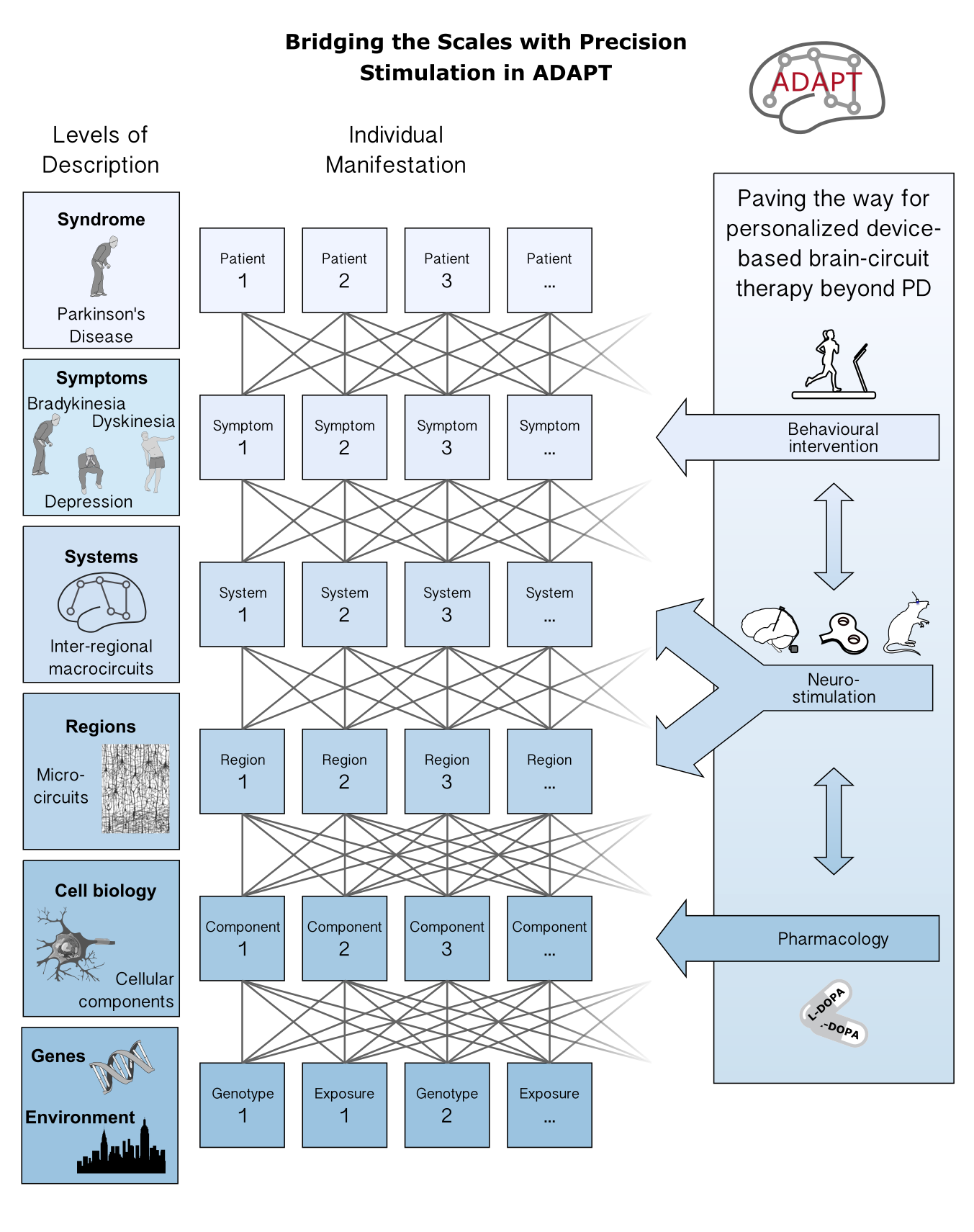
Many members of the Movement Disorders group are part of the ADAPT-PD project. Here, we investigate dysfunctional circuit dynamics in cortico-basal ganglia projections in PD with the aim to “normalize” them with brain stimulation techniques. ADAPT-PD is a collaborative international, translational and multi-modal project involving different techniques (invasive and non-invasive recordings as well as brain stimulation) in animal models and PD patients. Detailed information can be found here.
People: Aino Lindroos Jensen, Amin Ghaderi Kangavari, Ann-Charlot Rughaven, David Meder, Gustaf Olsson, Hartwig Siebner, Jiahua Xu, Jonas Laugesen, Kentaro Kawamura, Lasse Christiansen, Laura Sakalauskaitéé, Mattias Rickhag, Mihai Atudorei, Sai, Sune Gram Thomsen, Vinooth, Yufei Song
Unravelling altered network dynamics in the mid-brain and striatum in Parkinson’s disease with ultra-high field MRI
In this project, we use ultra-high field (7T) functional and structural MRI to map the individual degree and spatial pattern of neurodegeneration in PD patients and relate it to the patient’s motor and non-motor symptoms. We focus on two neurotransmitter systems, the dopaminergic and the noradrenergic system. The large majority of dopamine neurons are situated in two midbrain nuclei, the ventral tegmental area (VTA) and the substantia nigra pars compacta (SNc). Using high resolution imaging, we can show that dopaminergic neuron loss and iron accumulation within the SNc show an overlapping, but also spatially distinct disease pattern (Madelung et al. 2025).
The locus coeruleus (LC), a small nucleus in the brain stem, is the main source of noradrenergic projection neurons and it is also heavily affected by neurodegeneration in PD. Noradrenaline lies at the core of eliciting an arousal response to novel and surprising events and has a tight coupling to the sympathetic nervous system. We can show that the LC does not degenerate uniformly and that cell death in different parts of the structure correlate with different non-motor symptoms (Madelung et al. 2022).
People: David Meder, Hartwig Siebner, Sune Gram Thomsen, Weijian Liu
7T-RBD
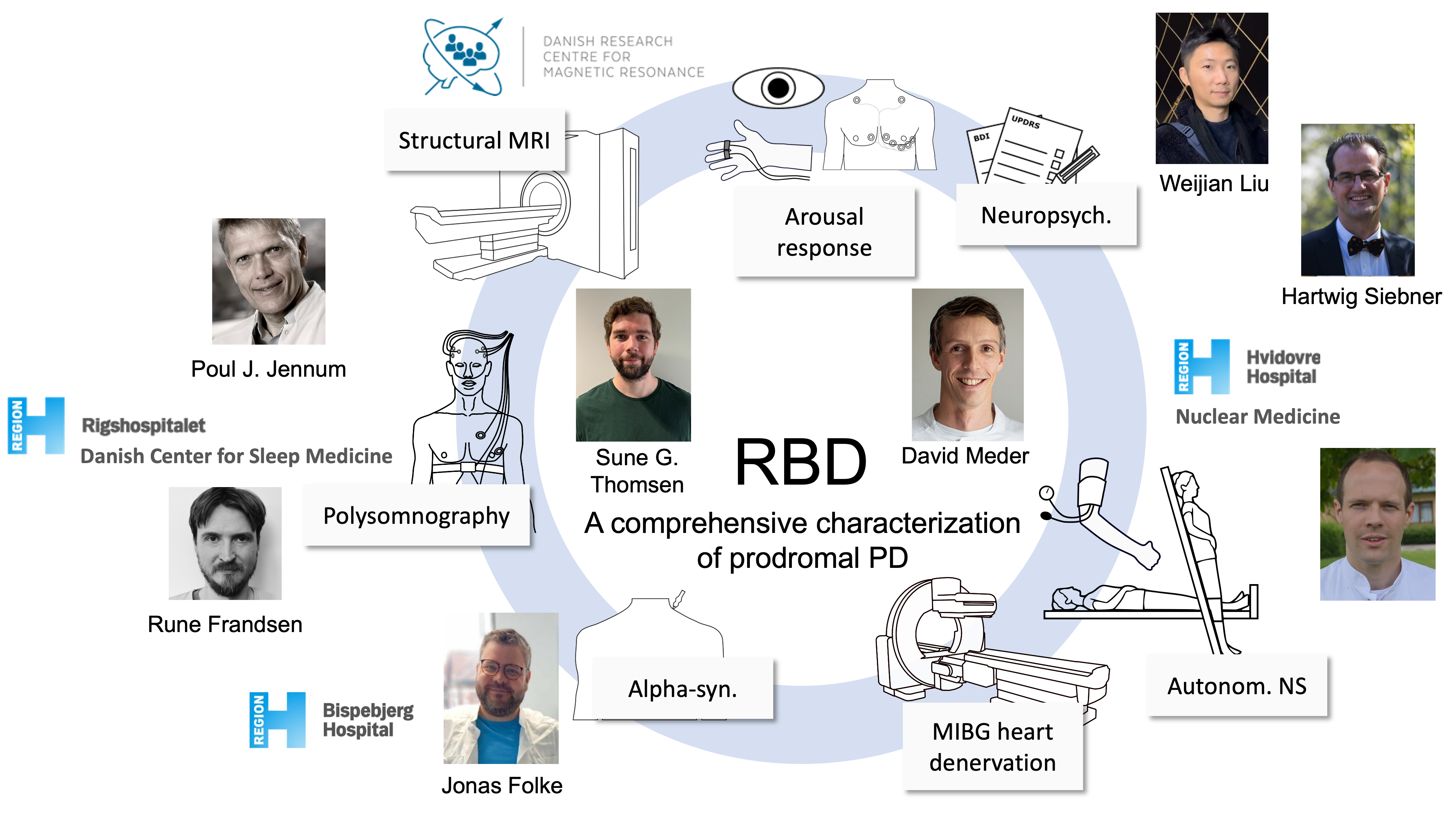
In this project, we extend the structural imaging to a cohort of patients with REM-sleep behavior disorder (RBD). Patients with this disorder have a high probability of developing an alpha-synucleopathy such as Parkinson’s disease, Lewy-body dementia or multiple system atrophy. Since the conversion rate to PD is considerably higher than the other synucleinopathies, RBD is often considered a prodromal state of PD. In this collaborative project, we aim to achieve a comprehensive characterization of these patients, combining our structural ultra-high field scanning with neuropsychological and motor performance measures, detailed sleep assessment with polysomnography from the Danish Center for Sleep Medicine at Rigshospitalet Glostrup, a thorough assesment of the autnomic nervous system at the Department for Nuclear Medicine at Hvidovre Hospital and finally measures of alphy-synuclein via blood-samples and skin biopsies.
People: Sune Gram Thomsen, David Meder, Hartwig Siebner
OPD - Optimism and Pessimism in the Dopamine System in PD
In the OPD project, we test a novel theory suggesting a hitherto unkown diversity in dopaminergic signaling. The main aim is to investigate whether the healthy human brain shows a diversity of optimistic and pessimistic reward signals and whether changes in this distribution in PD can provide mechanistic insights into the cause of symptoms. In order to address these questions, we will apply a unique combination of theory and methods on multimodal imaging and behavioural data acquired in healthy participants and PD patients. Using the fMRI signal in dopaminergic regions as proxy for regional dopaminergic signalling, we will probe reward and movement related activity in VTA/substantia nigra pars compacta (SNc) and striatum with functional magnetic resonance imaging. We will derive functional topographic maps of optimism/pessimism by applying a novel neuroimaging analysis method to the fMRI data developed at the department. Observing which parameters of the model are changed in the disease can then lead to a mechanistic understanding of how the disease affects brain function, leading to different symptoms.
People: Ditte Høier Frantzen, Amin Ghaderi Kangavari, Sune Gram Thomsen, Frederik William Løwe Lassen, Ester Bech Lambertsen, David Meder, Hartwig Siebner
Selected Publications
(Atudorei et al., 2025; Chen Thomsen et al., 2024; del Agua Villa et al., 2024; Haagensen et al., 2020; Herz et al., 2021; Lohse et al., 2023; Lund et al., 2026; Madelung et al., 2025, 2022; Meder et al., 2019)
Atudorei, M., del Agua Villa, C., Gether, U., Cenci, M.A., Siebner, H.R., Rickhag, M., 2025. Bilateral chemogenetic activation of intratelencephalic neurons in motor cortex reduces spontaneous locomotor activity in mice. Neurobiology of Disease 204, 106755. https://doi.org/10.1016/j.nbd.2024.106755
Chen Thomsen, B.L., Vinding, M.C., Meder, D., Marner, L., Løkkegaard, A., Siebner, H.R., 2024. Functional motor network abnormalities associated with levodopa-induced dyskinesia in Parkinson’s disease: A systematic review. NeuroImage: Clinical 44, 103705. https://doi.org/10.1016/j.nicl.2024.103705
del Agua Villa, C., Atudorei, M., Siebner, H.R., Rickhag, M., 2024. Pharmacological targeting of dopamine D1 or D2 receptors evokes a rapid-onset parkinsonian motor phenotype in mice. European Journal of Neuroscience 60, 7006–7024. https://doi.org/10.1111/ejn.16622
Haagensen, B.N., Herz, D.M., Meder, D., Madsen, K.H., Løkkegaard, A., Siebner, H.R., 2020. Linking brain activity during sequential gambling to impulse control in Parkinson’s disease. NeuroImage: Clinical 27, 102330. https://doi.org/10.1016/j.nicl.2020.102330
Herz, D.M., Meder, D., Camilleri, J.A., Eickhoff, S.B., Siebner, H.R., 2021. Brain Motor Network Changes in Parkinson’s Disease: Evidence from Meta-Analytic Modeling. Movement Disorders 36, 1180–1190. https://doi.org/https://doi.org/10.1002/mds.28468
Lohse, A., Løkkegaard, A., Siebner, H.R., Meder, D., 2023. Linking Impulsivity to Activity Levels in Pre-Supplementary Motor Area during Sequential Gambling. J. Neurosci. 43, 1414–1421. https://doi.org/10.1523/JNEUROSCI.1287-22.2023
Lund, A.E., Madelung, C.F., Madsen, K.H., Thomsen, B.L.C., Løkkegaard, A., Meder, D., Siebner, H.R., 2026. Blunted response of caudal locus coeruleus to arousing stimuli in Parkinson’s disease. Brain in press.
Madelung, C.F., Løkkegaard, A., Fuglsang, S.A., Marques, M.M., Boer, V.O., Madsen, K.H., Hejl, A.-M., Meder, D., Siebner, H.R., 2025. High-resolution mapping of substantia nigra in Parkinson’s disease using 7 tesla magnetic resonance imaging. npj Parkinsons Dis. 11, 1–12. https://doi.org/10.1038/s41531-025-00972-7
Madelung, C.F., Meder, D., Fuglsang, S.A., Marques, M.M., Boer, V.O., Madsen, K.H., Petersen, E.T., Hejl, A.-M., Løkkegaard, A., Siebner, H.R., 2022. Locus Coeruleus Shows a Spatial Pattern of Structural Disintegration in Parkinson’s Disease. Movement Disorders 37, 479–489. https://doi.org/10.1002/mds.28945
Meder, D., Herz, D.M., Rowe, J.B., Lehéricy, S., Siebner, H.R., 2019. The role of dopamine in the brain - lessons learned from Parkinson’s disease. NeuroImage, Mapping diseased brains 190, 79–93. https://doi.org/10.1016/j.neuroimage.2018.11.021
Funding
2025
Research grant from the Lundbeck Foundation (Experiment grant) for uncovering the underlying components of Bradykinesia (PI David Meder) – 2.300.000 DKK
2024
Research grant from the Danish Parkinson Association for assessing 7-tesla MRI as biomarker in REM-sleep behavior disorder (PI David Meder) – 264,000 DKK
Research grant from the Lundbeck Foundation (LFIN Seed grant) for testing the effect of vagus nerve stimulation on the arousal response (PI Daivd Meder) - 112,500
2021
Research grant from the Lundbeck Foundation (Collaborative grant) for ADAptive and Precise Targeting of cortex-basal ganglia circuits in Parkinson´s Disease (ADAPT-PD) (PI Hartwig Siebner) – 35.000.000 DKK
Research grant from Independent Research Fund Denmark (Sapere Aude) - Optimism and Pessimism in the Dopamine System in PD (PI David Meder) – 4.430.000 DKK